Pioneering the Future
Stories of Discovery & Innovation


Building the Body
May 17, 2022
Biologists who investigate how the intricate architecture of the human body takes shape are witness to extraordinary transformations, from an abrupt reprogramming that changes the way genes are used in a developing embryo to the emergence of fully functional organs and precisely wired neural circuits. Their discoveries offer insights into how and why development sometimes goes awry, as well as how a carefully structured body plan can deteriorate with age or disease—ultimately suggesting strategies for preventing or correcting such problems.
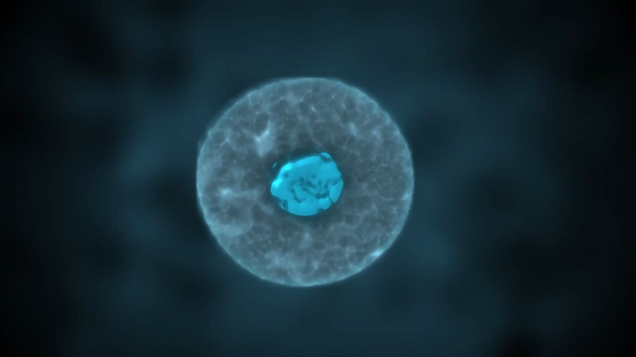

In the early stages of development, when a fertilized egg transforms into a small cluster of embryonic cells, major changes unfold. DNA and its associated packaging proteins are remodeled and reprogrammed so that the cells of the growing embryo can diversify and take on the many different roles needed to build a body.
At the Huntsman Cancer Institute at the University of Utah, Brad Cairns, PhD, and his team study how embryonic cells acquire this extraordinary developmental potential. They have identified a protein called DUX4 that orchestrates extensive reprogramming of developmental potential by activating hundreds of genes early in development. The discovery may lead to new insights into the causes of infertility and pregnancy loss. Additionally, it could help researchers learn more about facioscapulohumeral muscular dystrophy, a degenerative muscle disease caused by improper expression of the DUX4 gene in muscle cells, where it is normally silent.

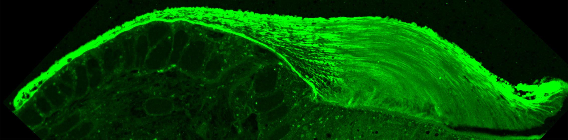
To help us hear, sensory cells in the inner ear rely on a tiny gelatinous structure called the tectorial membrane, which propagates and amplifies sound waves. The thin membrane is built before birth, and if its complex structure is improperly assembled, hearing will be impaired.
Neurobiologist Sungjin Park, PhD, and colleagues have found that the tectorial membrane, which is built not of cells but of extracellular materials like collagen, takes shape layer by layer. Park likens this sequential process of building the membrane to 3D printing. During assembly, the growing membrane is tethered to the cells that produce the materials from which it is being built. Once the matrix is assembled on the cell surface, it is released from its tether and a new layer forms underneath. The team’s findings not only clarify how this specialized structure forms in the ear, but also sheds light on the assembly of extracellular structures throughout the body.
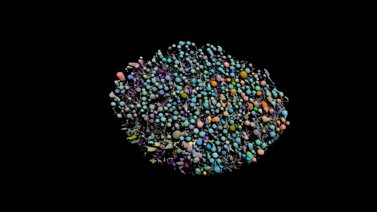

Millions of neurons in the retina of the eye work together to detect light and images. Their communications depend on intricately interconnected circuits that must be wired up properly so they can relay the right information to the brain.
Neuroscientist Bryan Jones, PhD, and his colleagues from the John A. Moran Eye Center used electron microscopes to visualize and map the neural circuitry in the eye with extraordinary detail. After charting the neural connections in a healthy eye, the team went on to show how these precisely wired circuits can deteriorate in neurodegenerative conditions like macular eye disease and diabetic retinopathy. Rewiring retinal degenerations are responsible for severe vision impairment or blindness in more than 2.4 million Americans. The findings could help researchers control retinal remodeling to slow or even reverse this type of vision loss.


Pioneering the Future: Stories of Discovery & Innovation at University of Utah Health
Special thanks to Wes Sundquist and Alfred Cheung for their work to compile the discoveries and innovations that make this series possible.
Written by: Jennifer Michalowski
Editing by: Julie Kiefer
Layout and Design by: Kyle Wheeler
Production Supervision: Abby Rooney, Julie Kiefer, Kyle Wheeler
Supported by: Will Dere, Chris Hill, Amy Tanner